Retina biomarkers identified 7 years before clinical diagnosis of Parkinson's disease
Researchers have identified markers that indicate the presence of Parkinson’s disease in patients an average of 7 years before the disease presents clinically.


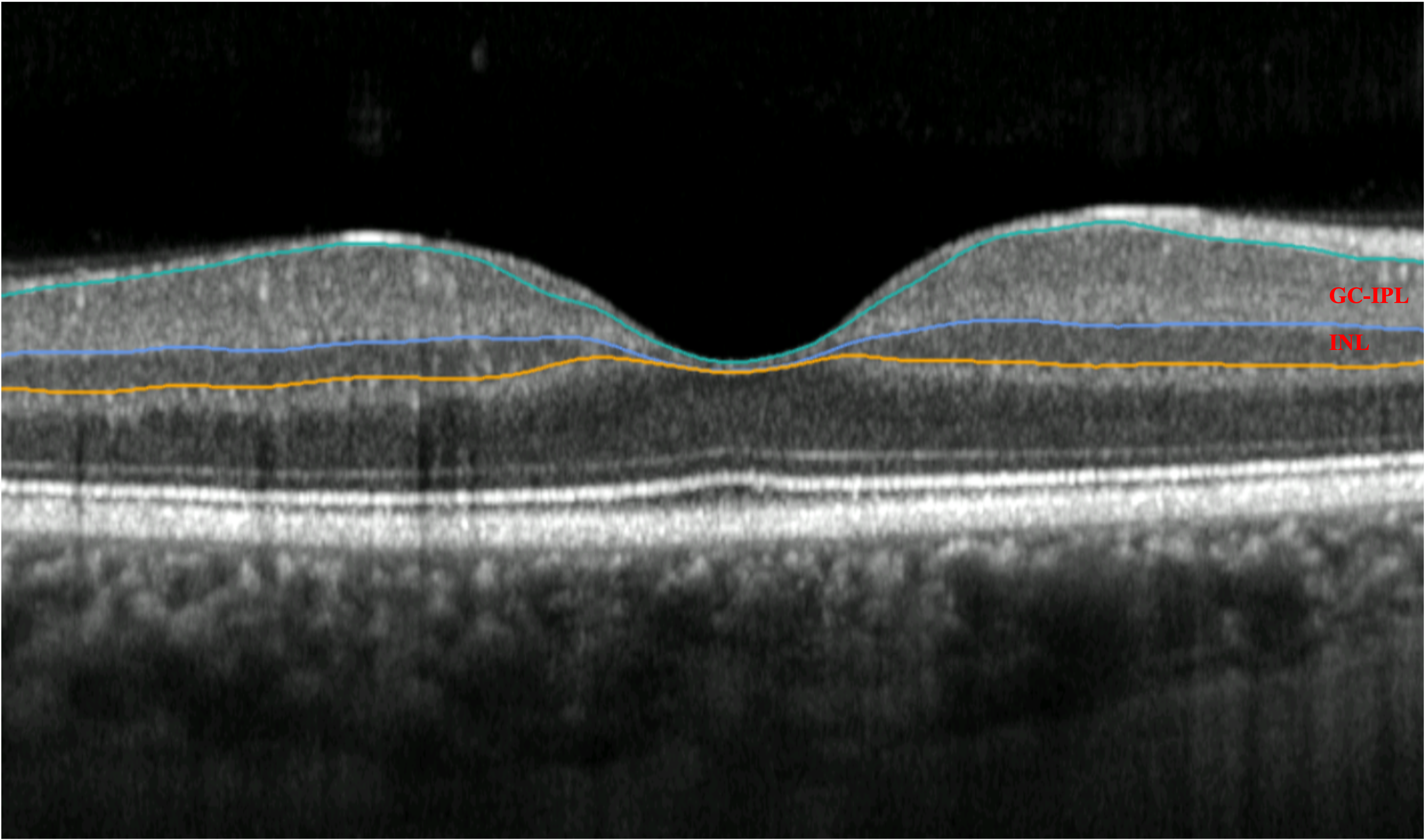
Image courtesy of the researchers
Researchers from Moorfields Eye Hospital and UCL Institute of Ophthalmology, London, have identified markers that indicate the presence of Parkinson’s disease in patients an average of 7 years before the disease presents clinically.
This is the first time anyone has shown these findings several years before diagnosis, and these results were made possible by the largest study to date on retinal imaging in Parkinson’s disease, according to the lead researchers Siegfried Wagner and Pearse Keane. They published their results in Neurology.
According to a press release, the study identified markers of Parkinson’s in eye scans with the help of artificial intelligence. Thescans taken from a simple optical coherence tomography (OCT) instrument, available to most ophthalmologists and optometrists.

")
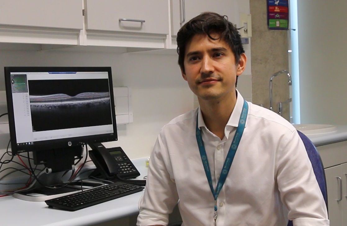
Siegfried Wagner, lead researcher with eye scan. (Image courtesy of Siegfried Wagner)
The authors commented that it seems most neurologic and cardiovascular diseases can be identified in the same way. This is part of an exciting new field called Oculomics, ie, using the eye as a window to the body to identify signs of disease no one would associate with it.
According to the press release, analysis of the AlzEye dataset was repeated using the wider UK Biobank database (healthy volunteers), which replicated the discoveries. The use of these two large powerful datasets has enabled the team to identify these subtle markers, even though Parkinson’s disease has a relatively low prevalence (0.1-0.2% of the population). Generation of the AlzEye dataset was enabled by INSIGHT, the world's largest database of retinal images and associated clinical data.
The use of eye scans has previously revealed signs of other neurodegenerative conditions, including Alzheimer’s, multiple sclerosis, and schizophrenia, in this emerging research field. The propensity to systemic disease such as elevated blood pressure, cardiovascular disease, and diabetes have also been identified through eye scans.
The work under discussion has involved collaboration between the National Institute of Health and Social Care (NIHR) Biomedical Research Centres at Moorfields Eye Hospital, University Hospital Birmingham, Great Ormond Street Hospital, Oxford University Hospital, University College Hospital London and the UCL Great Ormond Street Institute of Child Health.
Alistair Denniston, consultant ophthalmologist at University Hospitals Birmingham, professor at the University of Birmingham said: “This work demonstrates the potential for eye data, harnessed by the technology to pick up signs and changes too subtle for humans to see. We can now detect very early signs of Parkinson’s, opening up new possibilities for treatment.”
Dr. Wagner, clinical research fellow at Moorfields Eye Hospital, UCL Institute of Ophthalmology researcher and principal investigator for this and several other AlzEye studies, added: “I continue to be amazed by what we can discover through eye scans. While we are not yet ready to predict whether an individual will develop Parkinson’s, we hope that this method could soon become a pre-screening tool for people at risk of disease. Finding signs of a number of diseases before symptoms emerge means that, in the future, people could have the time to make lifestyle changes to prevent some conditions arising, and clinicians could delay the onset and impact of lifechanging neurodegenerative disorders.”
Louisa Wickham, Moorfields’ medical director, explained that “Increasing imaging across a wider population will have a huge impact on public health in the future, and will eventually lead to predictive analysis. OCT scans are more scalable, non-invasive, lower cost and quicker than brain scans for this purpose.”