Retina Society 2023: A modified endolaser instrument for chandelier scleral buckle procedures
A modified endolaser setup was found to be safe and efficient for use during chandelier scleral buckling.


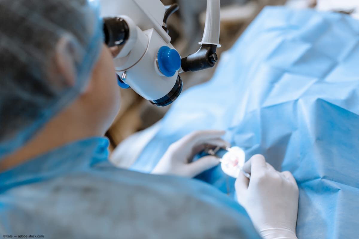
(Image Credit: ©Kate - Adobe.Stock.com)
Reviewed by Nilesh Raval, MD
A modified endolaser setup was found to be safe and efficient for use during chandelier scleral buckling. According to Nilesh Raval, MD,vitreoretinal surgery fellow at New York Eye and Ear Infirmary of Mount Sinai, New York, this instrument is successful in creating adequate retinopexy without disrupting the vitreous in young, phakic patients. Dr. Raval reported his findings during the 56th annual meeting of the Retina Society in New York.
He explained that laser or cryotherapy is generally used for retinopexy in scleral bucking procedures, however laser is typically done using indirect ophthalmoscopy which can be difficult in the setting of poor visualization, and cryotherapy portends an increasing risk of proliferative vitreoretinopathy. He and his colleagues modified an illuminated endolaser probe that facilitated safe endophotocoagulation with excellent visualization during chandelier-assisted scleral buckle surgery.
The modified endolaser probe was used in the treatment of 6 phakic patients with rhegmatogenous retinal detachments who underwent primary scleral buckling with 25-gauge chandelier endoillumination, external drainage, and laser endophotocoagulation using the novel, modified endolaser instrument.
Raval described that the instrument is comprised of an 18-gauge angiocatheter trimmed to create a sleeve that enshrouds the lighted endolaser probe tip, which shortens its reach beyond the extent of the trocar and prevents it from being inserted too deeply into the nonvitrectomized posterior segment.
The angiocatheter sleeve is trimmed so that after it is placed onto the endolaser probe shaft, the length of the exposed endolaser tip is equal to the length of a 25-gauge chandelier tip, which facilitates safe manipulation of the instrument in the vitreous cavity and without increasing the risk of iatrogenic retinal breaks. He pointed out that the laser power then can be titrated considering the distance from the retinal surface and the level of fundus pigmentation to optimize uptake.
The results indicated that the outcomes in all study patients were successful after primary scleral buckling was performed to repair retinal detachments using the modified endolaser instrument for intraoperative laser endophotocoagulation. All procedures were accomplished without any significant complications developing.
Raval and colleagues concluded that the modified endolaser setup was safe and efficient for use during chandelier scleral buckling.
“This instrument was successful in creating adequate retinopexy without disturbing the vitreous or contacting the lens in young, phakic patients. We recommend using this setup as an alternative to cryotherapy or indirect laser retinopexy during primary scleral buckling with chandelier-assisted endoillumination.”
Nilesh Raval, MD
E: nilesh.raval@mountsinai.org
Raval is an ophthalmology fellow at the New York Eye and Ear Infirmary of Mount Sinai, New York. He has no financial interest in this subject matter.