Handheld, spectrally encoded coherence tomography and reflectometry: Improving inclusive diagnostics with multimodality OCT
The investigators have developed a clinically robust HH-SECTR system, which has a modular aluminum probe design and micrometer-scale mechanical stability.
Reviewed by Jacob J. Watson and Rachel Hecht
Handheld optical coherence tomography (OCT) imaging and OCT angiography (OCTA) have the potential to improve diagnostic sensitivity for detecting eye diseases in patients who cannot have imaging done using a traditional benchtop examination. The engineers behind these technologies hope to improve point-of-care ophthalmic diagnostics and clinical imaging ergonomics for a variety of patient populations, including patients with limited mobility who cannot fixate themselves or sit upright as well as pediatric patients, such as infants born prematurely, who cannot cooperate for a traditional eye examination. Graduate students Jacob J. Watson and Rachel Hecht from the Department of Biomedical Engineering at Vanderbilt University in Nashville, Tennessee, reported the status of their research in handheld multimodality OCT at the Association for Research in Vision and Ophthalmology Annual Meeting 2023 in New Orleans, Louisiana.


Figure 1 HH-SECTR imaging probe. Top right: Modular aluminum HH-SECTR design. Bottom right: HH-SECTR being used for point-of-care retinal imaging. Left: HH-SECTR with enclosure and handgrip-integrated motorized focusing.
Image courtesy of Watson and Hecht

Commercially available handheld OCT/OCTA units are disadvantaged with slow imaging speeds, which can limit diagnostic sensitivity to fine features of the retina. Other research challenges associated with point-of-care eye imaging include motion correction, real-time and en faceretinal aiming, widefield imaging, and refractive correction. Refractive correction is of particular importance because manually correcting refractive errors to ensure optimal image quality can be clinically cumbersome during point-of-care eye imaging.
To overcome these challenges, the researchers are focused on developing a handheld spectrally encoded coherence tomography and reflectometry (HH-SECTR) system (Figure 1), which integrates OCT with complementary high-speed, real-time, en faceimaging.


Figure 2 Retinal imaging performed with handheld spectrally encoded coherence tomography and reflectometry. Left: Real-time, en face spectrally encoded reflectometry image. Center: En face OCT projection. Right (red box) and bottom (blue box): Corresponding OCT cross sections.
Image courtesy of Watson and Hecht
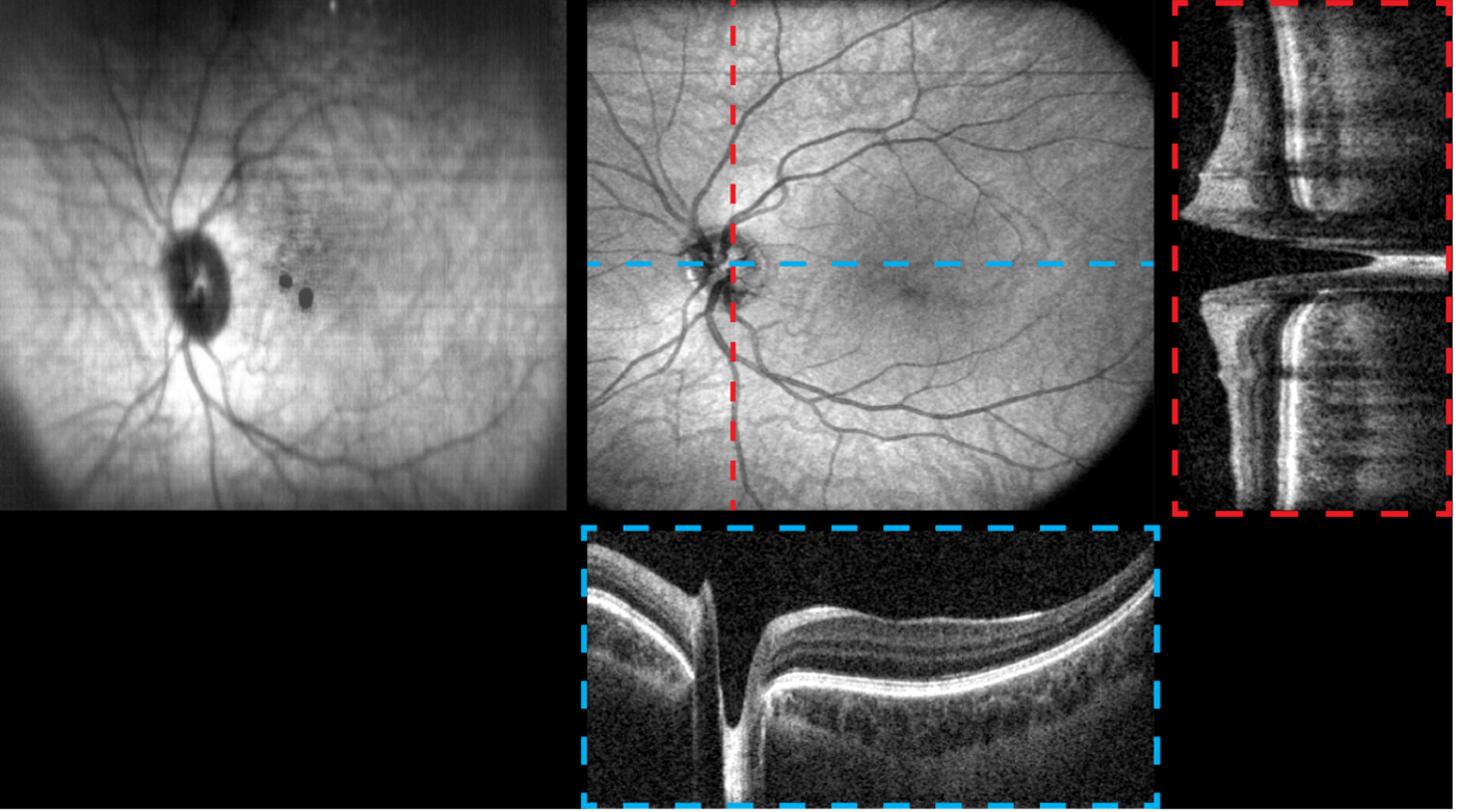
This allows the photographer to simultaneously see a cross section of the retina using OCT and a complementary top-down or en face view of the retina in real time (Figure 2). The researchers integrated a mechanical focus adjustment system into their handheld tool to make refractive correction more ergonomic to the photographer. They are also working to ensure that HH-SECTR has a robust form factor for translation into the clinic.
How this technology works
SECTR uses spatiotemporally and coregistered spectrally encoded reflectometry (SER) and OCT for simultaneous en faceand volumetric imaging. “We have improved our SER optics and imaging hardware to provide a better real-time en faceview from HH-SECTR. This will make aiming at different retinal features easier for the photographers and help our ability to correct motion artifacts that occur during handheld imaging,” Watson and Hecht said. They have also integrated a motorized focus adjustment system into HH-SECTR to enable ergonomic and high-quality imaging in patients without emmetropia. “This allows the photographer to maintain alignment of the handheld [system] to the pupil and retina while they adjust focus to get the best image quality.”
“We are also prototyping HH-SECTR out of aluminum using computer numerical control machining,” Watson and Hecht said. A modular aluminum handheld design ensures a reproducible, lightweight, and robust form factor for use in the clinic. The researchers also used structural analysis methods, such as finite element analysis, to assess the mechanical stability of their handheld system. “High mechanical stability lets us create the most lightweight handheld design [but] maintain optical alignment to ensure the best image quality during clinical use. Our current analyses indicate that the handheld [system] has micrometer-scale mechanical stability and weighs around 2 lb,” Watson and Hecht remarked.
Takeaways from this research
The investigators have developed a clinically robust HH-SECTR system, which has a modular aluminum probe design and micrometer-scale mechanical stability. They have improved the real-time en face imaging capabilities of SER to facilitate better handheld aiming at retinal features of interest. HH-SECTR also includes motorized focus adjustment capabilities, which can ergonomically correct –10 to +10 diopter of refractive error.
Watson and Hecht concluded, “We have demonstrated motorized focusing and retinal imaging with ournHH-SECTR system. Our next developments are to enable pediatric imaging and hands-free focus tracking. We predict these improvements will benefit the utility and clinical translation of HH-SECTR and ultimately improve point-of-care ophthalmic diagnostics.” •
Jacob J. Watson
E: jacob.j.watson@vanderbilt.edu
Watson is a graduate student from the Department of Biomedical Engineering at Vanderbilt University in Nashville, Tennessee. He has no financial interest in this subject matter.
Rachel Hecht
E: rachel.hecht@vanderbilt.edu
Hecht is a graduate student from the Department of Biomedical Engineering at Vanderbilt University in Nashville, Tennessee. She has no financial interest in this subject matter.