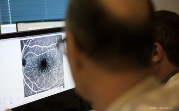
3D choroidal vessel imaging offers new insights into retinal diseases
Retina specialist Jay Chhablani, MD, discusses 3D choroidal vessel segmentation, uncovering critical insights into macular degeneration, diabetic retinopathy, and potential systemic disease connections using advanced OCT technology.
Jay Chhablani, MD, from the University of Pittsburgh Medical Center's Vision Institute discusses 3D choroidal vessel segmentation technology. Using advanced Plex elite Zeiss swept-source OCT imaging, his team has created a new tool to visualize and analyze Haller vessels in 3 dimensions – overcoming previous limitations of 2D imaging.
"We have been focused on choroid for more than a decade. Our team, along with machine learning engineers and clinical research fellows, have been working on choroid over the last few years, we have reported multiple choroidal biomarkers," Chhablani said.
The research focuses on examining choroidal vessel changes across multiple eye conditions, including
"So the key learning points, what we had was that as the disease starts in macular degeneration, these choroidal vessels start dilating, and the inter vessel distance starts coming down," Chhablani said. "And we saw this happening starting at very early stage to all the way to the...late AMD, including both geographic atrophy, as well as wet macular degeneration. We also compared the two eyes of the same patient, having one eye dry and one eye wet, and again, we saw that there was difference between these two eyes."
A particularly significant discovery is that choroidal vessels can change even when other retinal structures remain unchanged, suggesting the choroid plays a crucial role in disease mechanisms. By tracking early-stage AMD patients over a year, Chhablani's team observed that choroidal vessels were the only structures exhibiting modifications.
"Talking about central serous chorioretinopathy, we saw that as the disease is getting more chronic, these choroidal vessels are becoming much larger, the inter vessel distance is coming down, and that is where probably will explain the pachychoroid mechanism," Chhablani said.
"Talking about diabetic retinopathy, we saw that, in contrary to AMD and CSC, the vessel diameter is actually coming down, and that something which we understand that this has also been reported in histological studies of diabetic retinopathy patients. So we are yet to learn that how, what is the impact of proliferative diabetic retinopathy, as well as laser. And when we really think about macular degeneration over the most recent study, which we have submitted for review, is that when we picked up our early macular degeneration cases, followed them up for a year, and we saw that the choroidal vessels were the only changes which happened over 1 year – [the] rest of the retina looked absolutely same."
The implications extend beyond ophthalmology, with potential connections to systemic diseases like cardiovascular and kidney conditions. This is based on the understanding that choroidal vasculature is intimately linked to systemic vascular networks.
Technological advancements in OCT imaging have been instrumental in this research. The progression from time-domain to swept-source OCT, combined with wide-field imaging capabilities, has dramatically improved diagnostic precision and patient management. Chhablani emphasizes that these innovations allow for more comprehensive, earlier interventions in treating eye diseases.
Looking forward, the research aims to expand choroidal analysis across various ocular and systemic diseases, leveraging machine learning and advanced imaging techniques. This approach represents a significant step towards personalized, detailed understanding of vascular changes in health and disease.
The work underscores the importance of technological innovation in medical research, demonstrating how sophisticated imaging and analytical tools can unlock new insights into complex biological systems.
Newsletter
Keep your retina practice on the forefront—subscribe for expert analysis and emerging trends in retinal disease management.


