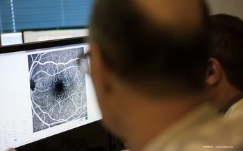
Q&A: Alan J. Franklin, MD, PHD, speaks to the use of intraoperative fluorescein angiography
Alan J. Franklin, MD, PhD, spoke with Modern Retina at Retina World Congress 2025. This meeting is taking place in Fort Lauderdale, Florida, from May 8-11, 2025.
In this conversation, Franklin discussed a research study on intraoperative fluorescein angiography in surgical procedures, highlighting significant advancements in surgical visualization and patient outcomes.
The following transcript has been edited lightly for clarity.
Modern Retina: You gave a presentation at Retina World Congress 2025 titled, Intraoperative fluorescein angiography for retinal vascular disease. What would you say are the key takeaways from this?
Alan J. Franklin, MD, PhD: First of all, I'd like to thank my co-authors. They were their pleasure to work with, and we've all contributed a lot to the to the data. Our highlights of the data is on intraoperative fluorescein angiography, and we're able to show that, based on the signals we see with fluorescein angiography intraoperatively, if we laser areas of fluorescein leakage or occult neovascularization, it reduces the post-operative vitreous hemorrhage rate to a really low rate, and also improves visual acuity in the first 3 months post-operatively.
MR: In your time as an ophthalmologist, how has advancements in imaging changed?
Franklin: Intraoperative fluorescein angiography was initially invented by Dr Charles, who's invented lots of stuff. With the operating microscopes, at the time, it was a little difficult to get good wide-angle visualization. Then researchers developed the technique for heads-up surgery, and then [ophthalmologists] streamlined it so that it's fairly easy to do with the retinal machine and the heads-up display. The pictures are very compelling, or the video is very compelling. But the question is, it's one thing to have nice pictures, it's another thing if the information helps your patients out. So that was kind of the impetus of the study. We thought we saw less vitreous hemorrhage in our patients after surgery. Then we said, well, let's get a group and look at people that have had the angiography and people that haven't. Let's formally look at it.
MR: As you look to the future of ophthalmology, where do you hope the industry goes?
Franklin: When I did my fellowship training a while ago, 25 years ago or so, even then, my mentors would say, focus on the image, the difference between the fellows and the attendings, where the attendings could show the vitreous better and show the pathology better with the light sources. It was really true. So now that we have better imaging, better visualization with the digital systems, we see the tissue better and what you can see better. You can operate more precisely with.
Newsletter
Keep your retina practice on the forefront—subscribe for expert analysis and emerging trends in retinal disease management.


