
|Articles|February 6, 2019
UWF imaging contributes to earlier disease detection
Author(s)Rishi P. Singh, MD, FASRS
New research confirms agreement with 7 standard field imaging
Advertisement
The body of research supporting the value of ultra-widefield (UWF) imaging is considerable and continuously growing. As a result, UWF is fast becoming the standard of care for retinal vascular disorders especially diabetic retinopathy.
The management of diabetic retinopathy (DR) presents a formidable and growing challenge to the ophthalmic community. According to the U.S. Centers for Disease Control (CDC), more than 30 million Americans (9.4% of the U.S. population) are diabetic,1 a number that is predicted to rise by 54% to 54.9 million by 2030.2 As a result, DR and other diabetic eye diseases are projected to follow a similar trend.3
DR is a silent disease that can manifest initially with few if any symptoms. While 40 to 45% of Americans diagnosed with diabetes are affected, only about half of these patients are aware they have diabetic eye disease.4 Consequently, many go untreated for far too long, losing vision and in some cases, going blind.
Thus, early detection particularly of those whose disease is most likely to progress, is critical for successful disease management.
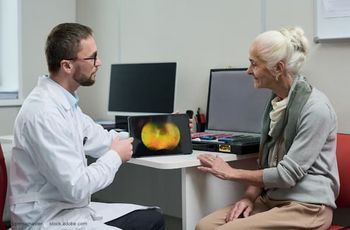
In my experience, the addition of ultra-widefield (UWF) imaging to screening and evaluation protocols offers advantages that improve our ability to diagnose earlier and treat more effectively. In recent decades, the standard for evaluating DR disease severity has been Early Treatment Diabetic Retinopathy Study (ETDRS) photography.
These 35-mm color images, comprised of 7 stereoscopic pairs of photographs per eye (ETDRS 7 standard fields), are assessed using the extended modified Airlie House classification system, which evaluates the location and degree of retinal lesions in the posterior pole.5
The ETDRS 7 standard fields includes the central posterior 90° of the retina, which equates to about 30% of the entire retina surface. For years this has been the gold standard for identifying vascular pathology in DR.
However, relying solely on this limited field of view means we risk missing a pathology present in the periphery that may contribute to the progression and outcome of the disease. DR studies have shown that pathology often exists outside the ETDRS 7 standard fields and in some cases, peripheral pathology is associated with greater disease severity and higher risk of disease progression.6–8
This has long been suspected, but recent research has begun to illuminate the role of peripheral pathology in early disease detection and determination of the risk of progression. Ischemia, which is an important factor in DR progression, may appear in the periphery first and has been associated with the presence of peripheral lesions on color images.9
One study found that predominantly peripheral lesions (PPLs) are present in up to 40% of patients with early nonproliferative diabetic retinopathy (NPDR) were linked to a nearly five-fold DR progression over 4 years.10
Defining UWF imaging
UWF images are defined as a single image capture that includes vortex ampullae in all four quadrants.11
Hundreds of published, peer-reviewed clinical studies have supported the value of the Optos 200° UWF based on its ability to improve screening for and identification of retinal disease.
In addition to the clinical and diagnostic benefits, because these images can be captured in less than ½ a second and without dilation, routine use of the technology can contribute to practice efficiency by facilitating assessment and documentation and allowing more patients to be screened in less time. The images are easily annotated, stored and shared, serving as a useful resource for making treatment decisions and referrals when necessary.
Other systems on the market capture varying degrees of the periphery using montaging techniques but they have not yet been widely adopted or validated versus gold-standard technologies.
UWF images also create a valuable tool for patient engagement and education. The ability to show patients the areas of concern or changes since a previous visit makes both their condition and your recommendations easier to explain and easier for the patient to understand.
Seeing the damage to their retina firsthand may even encourage compliance with treatment recommendations or inspire behavior modification, such as taking steps to improve blood glucose control.
UWF images = to ETDRS
While UWF imaging should not be considered a replacement for a dilated fundus exam, several published clinical studies have demonstrated its equivalence to ETDRS in the evaluation of DR severity. Recently, a large, multicenter, cross-sectional observational study conducted by the Diabetic Retinopathy Clinical Research Network (DRCRnet) found moderate to substantial agreement between ETDRS and Optos 200 degree UWF images.
Masked readers graded more than 700 subjects for DR level. When evaluated, the images agreed exactly in 435 eyes (59%) and were within one level in 96.9% (714 eyes). Additionally, the results indicated that UWF images were better for assessing DR level in 27% of eyes than ETDRS.
PPL were observed in 41% of these eyes and indicated increased DR severity by 2 steps or more in 11%. The authors concluded that these findings could support the use of UWF to evaluate DR severity in future clinical studies.12
UWF for telemedicine
UWF has also begun to be evaluated for its use in telemedicine environments given the ease of use, seamless integration with EMR and value of additional information captured. When implemented in one large telemedicine system, UWF detected double the amount of DR, reduced ungradable rates by up to 81%, due to the ability to easily image through small pupils and media opacity.13
Another study in a diabetic screening program found in eyes without diabetic retinopathy, approximately 20% may have ocular findings identified on UWF imaging.14 In our own Cleveland Clinic Executive Health Clinic, UWF imaging detected peripheral pathology in 18.4% of eyes not visualized by traditional small field imaging in a population of health screening subjects.15
Conclusion
The body of research supporting the value of UWF imaging technology is considerable and continuously growing; as a result, UWF is fast becoming the standard of care for retinal vascular disorders especially DR. In my practice, we rely heavily on both the clinical and practical advantages that this technology provides.
Given the evidence and my experience, I believe that we have a responsibility to adopt technology that has the potential to detect disease earlier, treat it more effectively and ultimately, provide better care to more of our patients.
Disclosures:
Rishi P. Singh, MD
E: drrishisingh@gmail.com
Dr. Singh is staff physician, Cole Eye Institute, Cleveland Clinic; medical director, Clinical Systems Office, Cleveland Clinic; and associate professor of ophthalmology, Case Western Reserve University. He is a consultant for Carl Zeiss Meditec and Optos.
References:
1. Centers for Disease Control and Prevention. National Diabetes Statistics Report, 2017. Atlanta, GA: Centers for Disease Control and Prevention, U.S. Dept of Health and Human Services; 2017.
2. Rowley WR, Bezold C, Arikan Y, Byrne E, Krohe S. Diabetes 2030: Insights from Yesterday, Today, and Future Trends. Popul Health Manag. 2017;20(1):6-12.
3. National Eye Institute. Projections for Diabetic Retinopathy (2010-2030-2050). https://nei.nih.gov/ eyedata/diabetic#5. Accessed January 14, 2019.
4. National Eye Institute. Facts about Diabetic Eye Disease. https://nei.nih.gov/health/diabetic/ retinopathy. Accessed January 14, 2019.
5. Early Treatment Diabetic Retinopathy Study Research Group. Grading diabetic retinopathy from stereoscopic color fundus photography-an extension of the modified Airlie House classification: ETDRS report number 10. Ophthalmology. 1991;98(5)(suppl): 823-833.
6. Wessel MM, Aaker GD, Parlitsis G, Cho M, D’Amico DJ, Kiss, S. Ultra-Wide-Field Angiography Improves the Detection and Classification of Diabetic Retinopathy. Retina. 2012; 32:785-791.
7. Silva PS, Cavallerano JD, Tolls D, Omar, A, Thakore K, Patel B, Sehizadeh M, Tolson AM, Sun JK, Aiello LP, Aiello PA. Potential Efficiency Benefits of Nonmydriatic Ultrawide Field Retinal Imaging in an Ocular Telehealth Diabetic Retinopathy Program. Diabetes Care. 2014;37(1):50-55.
8. Silva PS, Cavallerano JD, Sun JK, Soliman AZ, Aiello LM, Aiello LP. Peripheral Lesions Identified by Mydriatic Ultrawide Field Imaging: Distribution and Potential Impact on Diabetic Retionpathy Severity. Ophthalmology. 2013;120(12):2587-2595.
9. Silva PS, Dela Cruz AJ, Ledesma MG, vanHemert J, Radwan A, Cavallerano JD, Aiello LM, Sun JK, Aiello, LP. Diabetic Retinopathy Severity and Peripheral Lesions Are Associated with Nonperfusion on Ultrawide Field Angiography. Ophthalmology. 2015;122(12):2465-72.
10. Silva PS, Elmasry M, Pisig A, Aldairy Y, Van Hemert J, Fleming A, Sun, JK, Aiello LP. Automated Hemorrhage and Microaneurysm Counts on Ultra-widefield Images Predict Increased Risk of Diabetic Retinopathy Progression Over 4 Years. Paper presented at The Association for Research in Vision and Ophthalmology X Annual conference; 2018 Apr 29- May 3; Honolulu, HI.
11. Choudhry, N. Classification & Guidelines for Wide Field Imaging: Recommendations from the International Wide Field Imaging Study Group. Poster session presented at: 51st Annual Retina Society Meeting; 2018 Sept 12-15; San Francisco, CA.
12. Aiello LP, Odia I, Glassman AR, Melia M, Jampol LM, Bressler NM, Kiss, S, Silva PS, Wykoff CC, Sun JK. Comparison of Early Treatment Diabetic Retinopathy Study Standard 7-Field Imaging With UltrawideField Imaging for Determining Severity of Diabetic Retinopathy. JAMA Ophthalmol. 2019; 137(1):65-73.
13. Silva PS, Horton MB, Clary D, Lewis DG, Sun JK, Cavallerano JD, Aiello LP. Identification of Diabetic Retinopathy and Ungradable Image Rate with Ultrawide Field Imaging in a National Teleophthalmology Program. Ophthalmology. 2016; 123(6):1360-7.
14. Silva PS, Cavallerano JD, Haddad NM, Trolls D, Thakore K, Patel B, Sehizadeh M, Tolson AM, Sun JK, Aiello LP. Comparison of Nondiabetic Retinal Findings Identified With Nonmydriatic Fundus Photography vs Ultrawide Field Imaging in an Ocular Telehealth Program. JAMA Ophthalmol. 2016; 134(3):330-4.
15. Adhi M, Silva FQ, Lang R, Seballos R, Sukol R, Feinleib S, Singh RP. Non-Mydriatic Ultra-Widefield Imaging Compared With Single-Field Imaging in the Evaluation of Peripheral Retinal Pathology. Ophthalmic Surg Lasers Imaging Retina. 2017;48(12):962-968.
Advertisement
Related Content
Advertisement


Latest CME


Advertisement
Advertisement