
|Articles|June 8, 2018
Cutters or scissors for PDR?
Author(s)Vanessa Caceres
Advertisement
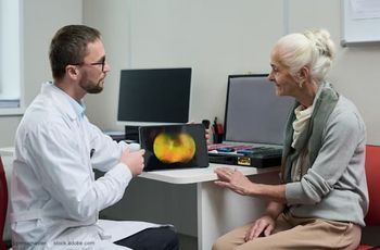
Retina surgeons should learn a variety of techniques to treat proliferative diabetic retinopathy (PDR) so they can select which one to use on a case-by-case basis, recommended Andre V. Gomes, MD, chief of the Retina Service, OSC Institute, Sao Paulo, Brazil.
Dr. Gomes addressed surgical approaches for PDR and the use of scissors versus cutters for PDR surgery.
The key steps of PDR surgery include:
" clearing the media
" removing fibrotic tissue and residual traction
" minimizing bleeding
" avoiding iatrogenic breaks and retinotomies
" reattaching the retina
" performing ERM and/or ILM peeling
" applying panretinal photocoagulation
" choosing a tamponade
"Patients should benefit from surgery both anatomically and functionally," he said. "They want to see better."
The past decade has brought with it an evolution in new technology and treatment options for retinal surgery in general and PDR surgery specifically, Dr. Gomes said.
This includes a new array of wide-angle viewing systems; new light sources, including "beautiful" chandelier lights so surgeons can work with both hands inside the eye; new "super high-speed" machines, a greater selection of gauges with larger openings at the very end of the tip, chromo vitrectomy, anti-vascular endothelial growth factor drugs that lead to less bleeding, and perfluoro-n-octane.
The technique choice for PDR surgery remains a topic of debate among retinal surgeons, Dr. Gomes said.
This includes the choice of probe only, the use of bimanual techniques, the choice of which gauge and which instrument to use, and whether phacoemulsification should be added to cases.
Dr. Gomes shared cases in which he used cutters or scissors for PDR. For instance, he shared one case in which he didn't feel comfortable using the cutter and had to use a pair of scissors. When a large retinal detachment is present, a blunt-tip scissors can be used to remove tissue in one piece, with no new breaks and no massive bleeding.
He shared another case in which he chose to use scissors at first and then used the cutter to trim part of the tissue.
"Without the help of a good pair of scissors, I couldn't finish this case in a safe manner," he said.
Dr. Gomes shared that when vessels get thicker and there is higher pressure as the surgeon goes deeper, it makes sense to cut them where they merge from the retina.
"That's what we do with scissors, especially when there is strong adherence on the optic disk," he said. "Laser to the periphery should be performed. There's less incidence of new bleeding and neovascular glaucoma."
After reviewing 320 of his organization's own cases, an algorithm he recommends is that if there is no retina detachment, especially if there is no large tabletop adherence to the retina, the surgeon is likely able to finish the case using only the cutter.
However, if there is large traction and there is retinal detachment and a thick tabletop-particularly if it extends to the periphery-surgeons may need to use a good pair of scissors.
"You can switch gauges in case you want a better pair of scissors inside the eye," he said.
Ultimately, surgeons should learn a variety of techniques so they can choose the appropriate one for each case, Dr. Gomes concluded.
Disclosures:
Andre V. Gomes, MD,
E:
This article was adapted from Dr. Gomes' presentation during Retina Subspecialty Day at the 2017 meeting of the American Academy of Ophthalmology. Dr. Gomes has no relevant financial disclosures.
Advertisement
Related Content
Advertisement


Latest CME


Advertisement
Advertisement