Bowtie artifacts: Clinical biomarkers for center-Involving DME?
A study of adult patients with diabetes found that bowtie-shaped polarization artifacts do not serve as reliable clinical biomarkers for center-involving diabetic macular edema. What might this mean for clinicians?
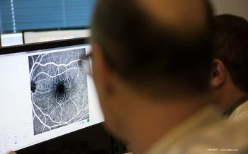
Nonconfocal ultra-widefield scanning laser ophthalmoscopy (SLO) images often contain foveal bowtie-shaped polarization artifacts.
A study of adult patients with diabetes found that these artifacts do not serve as reliable clinical biomarkers for center-involving diabetic macular edema (DME). Rather, they represent a preserved foveal Henle fiber layer structure, according to Radwan Ajlan, MBBCh, FRCS(C).
“Polarimetric imaging studies have demonstrated that the Henle fiber layer structure must be preserved for a foveal bowtie polarization pattern to occur.1,2 The prominence of these patterns declines with macular neovascularization, aging, and photoreceptor misalignment as well as with cornea and crystalline lens abnormalities.1-3
Ajlan; Luke Barnard, MS, and Martin Mainster, PhD, MD, FRCOphth, conducted a retrospective, observational, single-center cohort study of the foveal bowtie artifacts in nonconfocal ultra-widefield SLO images to see if that artifact was absent in patients with center-involving DME.
Seventy-eight patients (143 eyes; 42 men, 36 women; mean age, 64 years) with diabetes were included who had undergone spectral-domain optical coherence tomography (Spectralis, Heidelberg Engineering) and nonmydriatirc nonconfocal ultra-widefield SLO (Optos PLC, Nikon) examinations on the same day.
The investigators examined green, red, and pseudocolor images to determine if the bowtie-shaped foveal polarization artifact was present. The OCT images were reviewed for DME within 3,000 microns of the fovea.
Results
The authors reported their findings in Retina.4
Diabetic retinopathy was present in 116 of the 143 eyes, and DME was found in 50 eyes and was center-involved in 33 eyes.
Ajlan reported that the foveal bowtie polarization artifacts were absent when center-involving DME was present in all but one patient who had center-involving edema that was limited to the photoreceptor inner and outer segments. The artifacts also were not present in the eyes without center-involving DME in 30% of green light, 66% of red light, and 45% of pseudocolor SLO images.
“The presence of a bowtie pattern was negatively correlated with center-involving DME in all image subgroups (red light, 0.32 P = 0.0001; and green light and pseudocolor, -0.57 and -0.45, respectively; P<0.0001 for both comparisons),” Ajlan noted.
As a potential biomarker for center-involving DME in a diabetic population, he explained, the absence of a bowtie artifact has high specificity (99% for green light, 100% for red light, and 98% for pseudocolor SLO images, but poor sensitivity (49% for green light, 31% for red light, and 40% for pseudocolor SLO images).
The study concluded that “foveal bowtie-shaped polarization artifacts occur routinely in confocal ultra-widefield SLO images. Their presence indicates a preserved foveal Henle fiber layer structure. Contemporary nonconfocal ultra-widefield SLO images lack the sensitivity for their bowtie artifacts to serve as reliable biomarkers in screening for center-involving DME.”
References
Papay JA, Elsner AE. Near -infrared polarimetric imaging and changes associated with normative aging. J Opt Soc Am A Opt Image Sci Vis. 2018;35:1487-1495.
Weber A, Elsner AE, Miura M, et al. Relationship between foveal birefringence and visual acuity in neovascular age-related macular degeneration. Eye (Lond). 2007;21:353-361.
Bagga H, Greenfield DS, Knighton RW. Scanning laser polarimetry with variable corneal compensation: identification and correction for corneal birefringence in eyes with macular disease. Invest Ophthalmol Vis Sci. 2003;44: 1969-1976.
Ajlan RS, Barnard LR, Mainster MA. Nonconfocal ultra-widefield scanning laser ophthalmoscopy. Retina. 2020; 40-1374-1378.
Radwan Ajlan, MBBCh, FRCS(C)
E: rajlan@kumc.edu
Radwan Ajlan, MBBCh, FRCS(C), Luke Barnard, BS, and Martin Mainster, PhD, MD, FRCOphth are from the Department of Ophthalmology, University of Kansas School of Medicine, Prairie Village, KS. Ajlan and Barnard have no financial interest in this subject matter. Mainster is a consultant to Ocular Instruments, Inc.
Newsletter
Keep your retina practice on the forefront—subscribe for expert analysis and emerging trends in retinal disease management.


