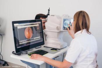
FLIO provides insight into autofluorescence lifetimes in AMD
Investigators find imaging method may be useful to monitor disease progression.
Reviewed by Jace Buxton.
Detecting age-related macular degeneration (AMD) as early as possible is the best strategy for preventing irreversible damage down the line. Fluorescence lifetime imaging ophthalmoscopy (FLIO), a noninvasive imaging method, may help delay progression by initiation of carotenoid supplementation, according to Jace Buxton, a medical student at the University of Utah School of Medicine in Salt Lake City.
Related:
FLIO’s characteristic pattern is a ring-shaped area of prolonged mean autofluorescence lifetimes around the macula. Buxton and colleagues conducted a study in which they investigated the changes in the autofluorescence lifetimes over time in patients with AMD to better understand disease progression and the responses to treatment.
Study and findings
The study evaluated 28 patients with a mean age of 73 years using a protype FLIO instrument (Heidelberg Engineering). Patients were followed for a mean of 17 months (range, 6-35). FLIO was performed to investigate a 30-degree retinal field centered on the fovea, Buxton explained.
Related:
Fundus autofluorescence was excited at 473 nm, and FLIO lifetimes were recorded in short (498-560 nm) and long (560-720 nm) spectral channels (SSC and LSC, respectively).
The investigators reported increases in the values obtained in both the SSC and LSCs over time. At baseline, the FLIO lifetimes in the outer ring of a standardized early treatment diabetic retinopathy study grid were 297±54 picoseconds (ps) and 405±58 ps, respectively, in the SSC and LSC. At the follow-up evaluation, the respective values the same areas were 306±59 ps and 411±57 ps, changes that reached significance in both spectral channels (P<0.01). The central area did not exhibit any significant changes in either spectral channel. The average 12-months prolongation of FLIO lifetimes in the outer ring was 7±20 ps and 7±13 ps in the SSCs and LSCs.
Related:
Buxton pointed out that the rate of progression varied among patients and even between eyes of the same patient. The investigators concluded that FLIO may be useful to monitor the progression of AMD as well as the effectiveness of treatment approaches. For example, in 3 patients treated with anti–vascular endothelial growth factor injections, there was a 10-ps annual decrease in the autofluorescence lifetimes.
Jace Buxton
E: Jace.Buxton@hsc.utah.edu
This article is adapted from Buxton’s presentation at the Association for Research in Vision and Ophthalmology 2021 virtual annual meeting. He has no financial interests related to this subject matter.
Related Content:
Newsletter
Keep your retina practice on the forefront—subscribe for expert analysis and emerging trends in retinal disease management.


