
Retinal vascular health: 4 insights ophthalmologists should keep top of mind
2024 is already bringing new research and insights to the forefront of retinal vein occlusion and providing new ways to consider retinal vascular health.


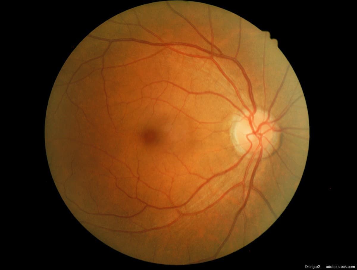
(Image credit: AdobeStock/singto2)
The vascular structure of the retina provides ophthalmologists and physicians with key insights on the health of a patient. This is why retinal imaging is key at every level of care, from routine optometric examines through the monitoring of condition progression in cases of retinal vein occlusion. With this in mind, here is why providers should keep in mind in 2024 and beyond for the retina’s vascular structure.
The retinal vein occlusion market will continue expanding.
The aging population along with this population’s escalating incidence of glaucoma and diabetes are key contributors to this expansion. This means that ophthalmologists will diagnose and treat a rising number of cases in the coming years. Luckily, advancements in diagnostics, imaging, and pharmaceutical innovations will help to meet the treatment needs of patients with RVO.
BALATON and COMINO phase 3 studies show retinal drying in RVO
At the 2024 Angiogenesis, Exudation, and Degeneration conference, Ramin Tadayoni, MD, PhD, head of ophthalmology at the Cité University in Paris, France, and president of EURETINA, presented 72-week data from 2 global Phase III studies, BALATON (NCT04740905) and COMINO (NCT04740931). These studies were evaluating faricimab (Vabysmo) in macular edema due to branch and central retinal vein occlusion (BRVO and CRVO).
In the news release about this presentation, Tadayoni said, “The sustained vision improvements and retinal drying seen up to 72 weeks reaffirm Vabysmo as an effective treatment for retinal vein occlusion.”
Read more about these trial results
Structural and vascular changes detected in RVO affected eyes and unaffected fellow eyes
In February of 2024, investigative ophthalmology & visual sciences published research from a team in Beijing that found Both eyes of patients with retinal vein occlusion (RVO), regardless of whether the eye is affected or is the unaffected fellow eye, have central and/or peripheral structural and vascular alterations in the retina and choroid. This research provides a basis for using ultra-widefield (UWF) swept-source OCT angiography enabled better visualization for a promising approach to more fully characterize the vascular alterations seen in RVO cases.
Read more about this study
Vascular abnormalities in the retina as may be biomarker for Alzheimer’s disease
Retinal vascular health is important not only for monitoring for eye diseases, but also as an early indicator for other conditions that may present elsewhere in the body. A recent study of changes in retinal structures suggested that significant vascular loss in cognitively healthy individuals before the onset of Alzheimer’s disease symptoms might be important biomarkers to identify pre-symptomatic Alzheimer’s disease.
Read more about this research
Newsletter
Keep your retina practice on the forefront—subscribe for expert analysis and emerging trends in retinal disease management.

 sBLA")


