RWC 2024: New technologies for navigated peripheral OCT of the vitreoretinal interface and retina
Modern Retina spoke with Paulo-Eduardo Stanga, MD, from The Retina Clinic London, London, United Kingdom at the 2024 Retina World Congress meeting.
Modern Retina spoke with Paulo-Eduardo Stanga, MD, from The Retina Clinic London, London, United Kingdom at the 2024 Retina World Congress meeting. He presented "New technologies for navigated peripheral OCT of the vitreoretinal interface and retina" at the event in Fort Lauderdale, Florida.
Video Transcript:
Editor's note: The below transcript has been lightly edited for clarity.
Paulo-Eduardo Stanga, MD:
I'm Professor Paulo-Eduardo Stanga. I'm a consultant retina surgeon. I specialize in both medical and surgical retina. I'm the director and lead surgeon at The Retina Clinic London in London, UK, and I'm a professor of ophthalmology at University College London.
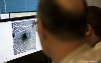
I would like to share with you our experiencing using a new technology for ultra widefield imaging. We are now relying on it [on a] daily basis with every single one of our patients, in the use of the Silverstone device from Optos. Every patient that comes to see us, before we see them, undergoes multimodal imaging, most importantly, ultra widefield imaging. So with a Silverstone device, what we are achieving is ultra widefield imaging and navigated peripheral OCT. So, we can either do central OCT, posterior pole OCT, or we can navigate the scan towards the periphery. All our technicians are trained to automatically scan any suspicious area in the retina, whether it is within the posterior pole, in the mid periphery, or in the periphery. But also very importantly, and something that's very exciting is that with this device, we can do also simultaneous either ultra widefield fluorescein angiography or ultra widefield indocyanine green angiography, and this with simultaneous navigated OCT, either central OCT or peripheral OCT. So, we are learning. We are learning about the association of different angiogeographic patterns with different patterns, cross sectional patterns, individual interface. For example, in diabetics, we can easily diagnose and differentiate airmass from neovascularization elsewhere in the cross-sectional scans. We can assess, for example, the presence of traction in different areas of the angiogram. And for example, diagnose peripheral vitreous cases very easily because of having the benefit of peripheral OCT. We can correlate ischemia with retinal thinning. We can correlate leakage with thickening of the retina. It's a learning, a learning experience we're going through now. This is the new frontier.
Newsletter
Keep your retina practice on the forefront—subscribe for expert analysis and emerging trends in retinal disease management.


