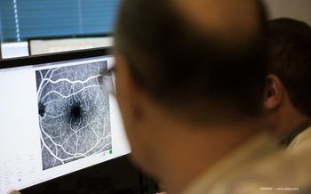
Tracking growth of ultra-widefield imaging in disease management
Ultra-widefield imaging has become an important component of a thorough eye exam and is taking on a larger role in retina and comprehensive ophthalmology.
Reviewed by Charles C. Wykoff, MD, PhD
As the popularity of ultra-widefield imaging grows and the technology becomes more commonplace, retinal specialists can use it to help with diagnosis and management decisions, said Charles C. Wykoff, MD, PhD.
“It’s a more complete view of the back of the eye,” said Dr. Wykoff, Retina Consultants of Houston, Houston. However, he added, it doesn’t replace a thorough clinical examination.
The advantage of using ultra-widefield imaging is its ability to capture more of the posterior segment than has been possible with traditional fundus photography.
The latest imaging technology (OptosAdvance, Optos) captures 200°, or 82%, of the retina in one image. The device has six imaging modes, including three-in-one color depth imaging, angiography, and autofluorescence. The device is also on a HIPAA-compliant, cloud-enabled platform so users can see images from any networked device.
“By definition, ultra-widefield images show a minimum of 80% of the retina and include key peripheral landmarks, such as the vortex ampullae,” said Carole McCallum, director of ophthalmology marketing, Optos.
Dr. Wykoff, who has used ultra-widefield imaging for more than six years, sees key advantages for the incorporation of ultra-widefield imaging into clinical practice. It can help guide the management of retinal vascular diseases including retinal venous occlusive disease and diabetic retinopathy (DR).
With ultra-widefield imaging, more pathology is often visible peripherally, Dr. Wykoff said.
Guiding treatment decisions
The technology may help guide treatment decisions. For example, a retina specialist may perform a clinical exam in a patient with DR, but then ultra-widefield angiography may detect a greater burden of disease.
“That can change prognosis and how one may manage the condition, such as with anti-vascular endothelial growth factor agents or retinal photocoagulation,” he said.
Dr. Wykoff also uses ultra-widefield imaging to document and track pathology, including retinal tears, retinal detachments, ocular tumors, and areas of inflammation.
“It’s helpful to objectively document areas of pathology with a single capture,” he said.
“Peripheral information seen in ultra-widefield color and fluorescein angiography have been shown to alter risk assessment and treatment plans in conditions such as diabetes and uveitis,” McCallum said.
Researchers are exploring ways to use ultra-widefield imaging, including for telemedicine screening, aging eye trends, and even for potential signs of Alzheimer’s disease. Clinicians are also analyzing the advantages of using ultra-widefield imaging to track DR in an ongoing, large, multicenter prospective study, Dr. Wykoff said.
Despite its potential, he cautioned that ultra-widefield imaging does not replace a dilated eye exam.
“The only way to see the entire retina in most eyes is to perform a scleral depressed examination in all directions,” he said.
Ultra-widefield imaging can be particularly limited in the inferior quadrants. he added.
Dr. Wykoff’s research involvement with ultra-widefield imaging includes work towards accounting for the warping inherent to ultra-widefield imaging1 as well as its application to clinical trials such as the WAVE trial.2
Going forward, Dr. Wykoff would like to see ultra-widefield imaging extend even further peripherally, particular to the ora seratta in all directions, and believes that integration of software algorithms for image interpretation will be used to quantify levels of pathology including DR severity levels in the near future.
Reference
1. Croft DE, van Hemert J, Wykoff CC et al. Precise montaging and metric quantification of retinal surface area from ultra-widefield fundus photography and fluorescein angiography. OSLI Retina. 2014 Jul-Aug;45:312-317.
2. Wykoff CC, Ou WC, Wang R, et al. Peripheral laser for recalcitrant macular edema owing to retinal vein occlusion: The WAVE trial. Ophthalmology. 2017;124:919-921.
Charles C. Wykoff, MD, PhD
E: ccwmd@houstonretina.com
Dr. Wykoff does not have any relevant financial interests to disclose.
Newsletter
Keep your retina practice on the forefront—subscribe for expert analysis and emerging trends in retinal disease management.


