|Articles|August 24, 2022
- Modern Retina Summer 2022
- Volume 2
- Issue 2
Achieving high-definition slit lamp imaging via smartphone mirroring
Author(s)Sunil Mamtora, MD
This affordable device allows the view through a slit lamp to be shared in real time or recorded, for teaching and reference purposes.
Advertisement
Special to Modern Retina™.
Learning how to use the slit lamp is not easy. I think as ophthalmologists we take this skill for granted and forget the journey we undertook to master basic examination techniques. I vividly remember sitting in the clinic during my ophthalmology rotations as a medical student and wishing I could see what the consultant was seeing.
When I was asked to have a look through the slit lamp, I often ended up nodding and smiling to avoid the embarrassment of admitting that I had no idea what I was looking at. (The patient had probably moved their eye and what the consultant had wanted me to see was no longer in the field of view.)
On rare occasions, an observer tube was connected to the slit lamp and, although this was somewhat useful, one had to be careful to not have it bang into one’s eye while crouching uncomfortably beside the slit lamp.
I had a revelation at the beginning of my ophthalmology residency some years ago when I witnessed the use of an anterior segment camera. Why not use this camera to teach people how to use slit lamps?
My consultants at the time seemed indifferent to this suggestion but one of my seniors agreed with me. So when the camera was not in use (the vast majority of the time), we used it in the clinic. It helped me to develop my clinical skills and I found it particularly useful for learning how to examine the cornea and recognize a huge variety of pathologies.
With the consent of the patients we saw, I made a video library, which I attached my notes to. Feeling vindicated, as well as passionate about the benefit of slit lamp cameras for ophthalmic education, I fought for a grant for teaching on slit lamps after securing a loaned device for a few months.
Positive feedback
The feedback from junior doctors, nursing colleagues, and medical students was overwhelmingly positive. We even managed to connect a virtual reality headset to a beam splitter/slit lamp camera with real-time video output to create an immersive viewing experience. This set-up was presented at the American Academy of Ophthalmology meeting in 2018.
We were successful in securing funding of almost $19,000 to purchase a MediWorks digital slit lamp, and I spent just over a year using this system before having to move on to a new hospital as part of my registrar rotations in southwest England. We made the most of the time we had and I set up a YouTube channel with the support of my friends and colleagues, amassing thousands of followers. Some videos have received almost a million views.
When I moved to my new rotation, I was disappointed to find that, rather unsurprisingly, there was no slit lamp camera. The overwhelmingly complex and high-effort process of applying for funding for a slit lamp camera system was too much to contemplate at that time.
I stumbled across the MicroREC optical system (Custom Surgical) through an advertisement on social media. The device is predominantly used to connect a smartphone to operating microscopes, to allow the user to record surgical videos on their phone. However, it can also connect to slit lamps.
I was lucky enough to have a beam splitter for a Haag-Streit BQ900 slit lamp, which allowed me to use the optical system. I initially combined it with an iPhone 8 and subsequently an iPhone 12 Pro Max, with incredible results.
Normally in a National Health Service hospital in Britain, one is lucky if there is 1 slit lamp with a camera system and even luckier if an ophthalmologist can have their clinic in the room it is located in. I had a 4K slit lamp camera system that I could keep in my bag and use on any slit lamp for less than $1250! I further enhanced the system by adding a diffuse illuminator that I purchased from Eye2Mobile.
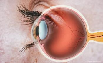
Slit lamp imaging is incredibly valuable and it amazes me that it is not seen as a fundamental part of the hospital eye service. Although diagrams in the notes are useful, there is no doubt in my mind that a photo is far more objective and useful when combined with a clinician’s impression of the patient’s condition.
Shorter learning curves
Teaching clinical skills is far easier with the use of a slit lamp camera system: using an Apple TV device I can “cast” a real-time, live view of the slit lamp from my iPhone to a computer monitor. I successfully taught a medical student how to perform a fundus examination in just 1 afternoon, a skill that often takes weeks if not months to learn.
I was also able to teach a nurse practitioner how to perform Goldmann applanation tonometry proficiently over 2 clinics. The practitioner had been sitting in on 1 glaucoma clinic a week over the previous 2 years to try to learn this skill, without success.
Being able to see what the student sees allows one to provide critical, accurate, and useful feedback (eg, that the Myers rings are too thick or that one needs to move closer). Indeed, alignment to the center of the cornea, adjustment of the tonometer, and so on are only possible when one can see what the student is seeing and vice versa.
With the backlog of patients as a result of the pandemic, we need to focus on how to teach basic clinical skills to junior doctors and allied health professionals in the most immersive and efficient way possible. Slit lamp camera systems or digital slit lamps are a low-hanging fruit that will enable the acquisition of this skill, and we have proved this in a small pilot study.1
Having an optical system that allows you to capture procedures while performing them democratizes digital ophthalmic imaging, not just regarding the slit lamp but also in terms of surgical training, for which camera systems for operating microscopes are often prohibitively expensive.
Smartphone cameras are incredible: the quality of photos and videos that can be captured from a mobile phone these days is staggering. The latest devices can capture video in up to 8K for breathtaking quality.
Additionally, with the apps that come with many ophthalmic devices now, one can optimize the videos or photos captured and export or share them in various formats, including a low file size, compressed version, which allows for quick distribution on social media. This removes the need to purchase an additional camera system, and having the videos on one’s own device means they can be shared with colleagues (with patients’ permission, of course) when advice is sought out or watched at home to hone surgical skills.
The ability to conduct remote slit lamp video consultations was something we found particularly beneficial during the peak of the pandemic. I hope that ophthalmologists will increasingly come to realize the importance of digital imaging in clinical ophthalmology. Maybe in the future we will laugh at how we used to make diagrams on paper notes with colored pencils while reviewing slit lamp videos on 8K TVs.
Sunil Mamtora, MBBS
E: s.p.mamtora@hotmail.com
Mamtora is an ophthalmology academic specialist registrar at Bristol Eye Hospital, England; chair of the Ophthalmologists in Training Group of the Royal College of Ophthalmologists; and host of the Eye to Eye podcast. He has no relevant financial disclosures.
Reference
1. Smith C. Teaching ophthalmology: mastering the slit lamp examination. Inspire Student Health Sciences Research Journal. Winter 2020. https://inspirestudentjournal.co.uk/wp-content/uploads/2020/10/Inspire-Student-Journal-Chanelle-Smith.pdf
Articles in this issue
almost 4 years ago
Gene therapy: Where the action is for retinal diseasesalmost 4 years ago
Practice perspective: Academic retinaalmost 4 years ago
Perspectives on intravenous bevacizumab repackaged for intravitreal useAdvertisement
Related Content
Advertisement

Latest CME


Advertisement
Advertisement