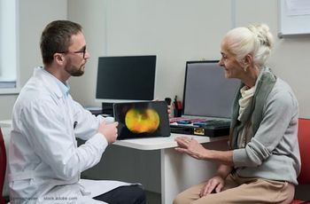
Heads-up surgery: Current advantages debatable, but system offers opportunities for the future
Retinal specialists discuss the role of “heads-up” surgery.
Take-home message: Retinal specialists discuss the role of “heads-up” surgery.
Reviewed by Steve T. Charles, MD, and Claus Eckardt, MD
Certain features of “heads-up surgery”, which is performed with a proprietary three-dimensional (3-D) visualization platform (TrueVision System), make it attractive new technology, agree Claus Eckardt, MD, and Steve Charles, MD.
The two retinal specialists, however, offer differing views on its most important advantages.
In heads-up surgery, 3-D cameras are attached to the operating microscope optics, and the surgeon views a stereo image of the surgical field on a flat panel LCD monitor with passive glasses.
Noting that he and his colleagues at Klinikum Frankfurt Höchst, Frankfurt, Germany, have performed more than 3,500 anterior and posterior segment surgical cases using the heads-up technology, Dr. Eckardt highlighted the high dynamic range of the 3-D cameras, the large display, ability to apply digital image processing to brighten the image, and comfort for the surgeon. In addition, he looks forward to augmented capabilities with future integration of systems such as overlay guidance and intraoperative OCT.
“There are enough reasons to believe that in the future, ophthalmic surgeons may no longer be looking through microscope eyepieces,” said Dr. Eckardt, professor of ophthalmology, Klinikum Frankfurt Höchst.
Although he does not consider the 3-D system to offer important ergonomic or visualization benefits, Dr. Charles is also looking ahead because he believes the technology will have value for opportunities provided through the incorporation of other imaging modalities requiring registration and tracking.
A true enthusiast’s view
In order to assess the utility, limitations, and benefits of the heads-up system for performing vitreoretinal surgery, Dr. Eckardt and colleagues undertook a study comparing it with the traditional approach [Retina. 2016;36:137-147]. Their assessments showed depth of field was similar using the heads-up system versus microscope oculars. Image resolution was about 30% less, although Dr. Eckardt suggested resolution with the 3-D system will be improved in the future. Furthermore, the larger image provided with the heads-up technology helps compensate for the difference, he said.
“It is not surprising that the image through the oculars’ has higher resolution considering the incredible resolution of the retina. In a visual field of 120°, theoretically more than 500 megapixels (MP) have to be filled in order to make them indistinguishable for our eyes,” he said.
“However, this is only valid with eye movements, and if the eyes are not moved to scan the whole image with the fovea, the image in the brain has a resolution of only 7 MP in the area of foveal fixation and 1 MP elsewhere.”
Although the 3-D camera system of the heads-up platform delivers a resolution of only 4 MP, Dr. Eckardt anticipates pixel density will steadily increase over time. Nevertheless, by showing an image displayed on his cell phone and on the larger screen of a tablet, Dr. Eckardt demonstrated that the viewer’s ability to discern detail is not all about resolution. Rather size also matters.
“We feel the large image displayed on the monitor of the heads-up system improves the depth perception and allows the surgeon to perform more precise surgery,” he said.
“In addition, it is possible to increase brightness with digital image processing. That can also mean better surgery and simultaneously allows reduction of the endoillumination level.”
Dr. Eckardt also touted the better ergonomics of operating with a heads-up view as well as the benefits it brings for teaching.
“Surgeons in training and others in the room can see exactly the same image the operating surgeon sees,” he said.
Alternative appraisal
Dr. Charles disagreed that the heads-up system provides better ergonomics, and he also noted the image suffers from a number of limitations. However, he believes the heads-up technology will definitely have a future role in vitreoretinal surgery because of the ability to display and overlay pre- and intra-operative imaging modalities such as en face OCT.
Discussing ergonomics, Dr. Charles said that problems with use of an operating microscope are an issue of the past thanks to the introduction of tilt oculars. On the other hand, use of the heads-up system creates an ergonomic problem because it requires surgeons to turn the head to the side to view the video monitor if the operating assistant has a direct view of the display. The set-up can also be reversed, but that situation “creates a nightmare for the assistant”, or the monitor can be placed far away at the patient’s feet. However, the latter arrangement typically does not allow unobstructed viewing, said Dr. Charles.
He noted that while the heads-up display found in aircraft cockpits is a valuable tool for pilots, it serves a different purpose than the surgical heads-up display, and so the image requirements are different. In aircraft, the pilot sees a live optical view through the cockpit windshield onto which there are superimposed a real time infrared image and/or a photo-realistically rendered 3-D terrain database, Dr. Charles explained. A full color 8 bit image would obstruct direct viewing out the windshield,” he said.
Commenting on the dynamic range of the LCD displays of current video systems, Dr. Charles said it falls orders of magnitude below that of the human eye, which has a dynamic range of 90 dB. There is opportunity for higher dynamic range using an OLED display, but that technology is costly and not currently used in the commercially available heads-up surgery system.
“CCD and even CMOS cameras have less dynamic range than the human eye and produce high dynamic range video only by using different gains on a sequence of frames,” he added.
The image appearing on the monitor of the heads-up surgery system also suffers from the limited color gamut of the LCD display. In addition, the appearance of colors and contrast are altered by a non-perpendicular viewing angle. As another practical issue, the image on the flat panel display is affected by reflected light, glare, dust, and fingerprints.
“With optical viewing using rubber eye shields, there is no extraneous light,” Dr. Charles said.
Improvement in image resolution with a heads-up system is limited by sensors and displays not the optical system of the operating microscope.
Dr. Charles also raised concern about the reliability of a heads-up surgery system, considering the possibility for failure with its various components, including a host personal computer, video boards, software, video display, cameras, and their respective power supplies.
“With the optical microscope, the lamp is the only failure mode, and that is easily addressed by keeping a spare lamp on hand. Otherwise, these systems are highly reliable, and even if there is loss of power, surgeons can still operate without power XY, zoom, and focus functions,” he said.
A heads-up display system is dependent on a computer and it is power supply as well as software.
Claus Eckardt, MD
E: Claus.Eckardt@KlinikumFrankfurt.de
Steve T. Charles, MD
E: scharles@att.net
This article is based on presentations given by Dr. Eckardt and Dr. Charles during Retina 2015. Dr. Eckardt was assigned to give a proponent’s view for the topic “Heads Up – No microscope is the future of vitreoretinal surgery.” Dr. Charles was assigned the opposing view and the negative arguments he presented do not necessarily reflect his personal opinions.
Dr. Charles and Dr. Eckardt have no relevant financial interests to disclose.
Newsletter
Keep your retina practice on the forefront—subscribe for expert analysis and emerging trends in retinal disease management.


