Leveraging noninvasive ophthalmic imaging for patients with Alzheimer disease and analyzing UK Biobank data
Tunde Peto, MD, PhD, discussed two of her presentations at EURETINA 2022: "UK Biobank retinal imaging grading: methodology, baseline characteristics and findings for common ocular diseases" and "Retinal phenotyping of different variants of Alzheimer’s disease using ultra-widefield imaging."
Video transcript
Caroline Richards: Thank you for joining me today, Tunde Peto, to discuss your presentations at the upcoming EURETINA conference. It's wonderful to have you.
So I understand that you will be presenting on UK Biobank grading data. Could you tell me a bit more about what the UK Biobank is and why it is important?
Tunde Peto, MD, PhD: So UK Biobank included about 500,000 participants from all areas of the UK. And the participants underwent a lot of questionnaires, testing, and also about 115,000 of them have ophthalmic data, and about 69,000 of them have color and OCT images.
We have manually graded to establish the ground truth for ophthalmic imaging for these patients. And I'm presenting the headlines for common blinding diseases, such as age-related macular degeneration, diabetes, and glaucoma. And we have linked these imaging data to hospital eye services data.
So there are some very exciting new findings that we will be—that will be coming out at EURETINA.
Richards: Oh, excellent. And what are the main learning points from your second presentation on imaging?
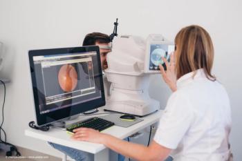
Peto: My second presentation on images is also very exciting because we have worked with a cohort of patients with Alzheimer's disease. And we have followed them up and we have very detailed Alzheimer and genetic recognition for them. And we found that patients with Alzheimer's disease present with characteristic changes in the back of the eye.
Why is it exciting? It's exciting because patients with Alzheimer's disease, they undergo a lot of very difficult for them and difficult for the health service treatment and also imaging such as MRI imaging, for example with some radioactive material.
And if we can choose eye imaging instead of some of the invasive imaging, then we will be able to make patients' and carers' lives much easier. And we will be able to follow these patients up with non-invasive techniques that would help both them, the carers and the society to make sure that they are appropriately seen but not overly burdened by the diagnostic and follow up investigations.
Richards: Oh, that sounds great. Thank you so much for filling me in on your two upcoming presentations at EURETINA. It was great to hear all of that information. Thank you very much.
Peto: Thank you.
Newsletter
Keep your retina practice on the forefront—subscribe for expert analysis and emerging trends in retinal disease management.


