Q&A: The increasing potential of ERG in diagnosing a wide range of eye conditions
Easier, more accessible functional studies have been made possible
The article was reviewed by Ruth Hamilton, PhD.
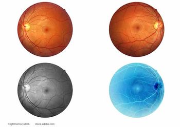
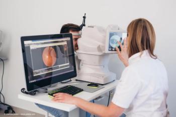
Electroretinography (ERG) has come a long way since its inception in the mid to late 1800s, when the notion of recording the eye’s electrical activity as a measure of its function was revolutionary.1 It was not until the 1900s that this technology was applied to patients in a clinically relevant manner (Figure 1).
Standardization of testing protocols by the International Society for Clinical Electrophysiology of Vision (ISCEV) enabled the development of clinical practice guidelines for the administration and interpretation of ERG so that patients would benefit from its diagnostic insights.
Today, the test has a wide range of applications such as inherited retinal conditions, medication-induced retinopathy, diabetic macular edema, vein occlusions, arteriosclerotic retinal damage, and Vitamin A deficiency, among others. I had the pleasure of speaking with Ruth Hamilton, PhD, the newly elected president of the ISCEV and the secretary of the British Society for Clinical Electrophysiology of Vision (BriSCEV), about the progress that has been made with ERG and its future potential.
JUSTUS: ERG is sometimes thought of as difficult to administer, difficult to interpret and irrelevant for most ocular complaints, and instead better suited for the retina clinic and research studies. How would you respond to individuals who feel this way?
HAMILTON: ERG has great promise to assist in clinical decision-making for patients with various ophthalmic conditions, but you are right that it is sometimes seen as a niche tool for use only by retina specialists and researchers. This reputation was likely born from the complexity of performing and interpreting the test, the length of time it can take to administer and the costs of devices. However, multiple studies suggest that ERG testing may be useful as a screening tool for bread-and-butter general ophthalmology diagnoses such as diabetic retinopathy (DR), glaucoma and hereditary diseases.2–5
The challenge of these conditions is that they are mostly asymptomatic until they are late-stage and have already caused irreversible disease that could have been avoided with early intervention. These conditions represent a significant international public health burden, with DR having a global prevalence of over 93 million people.6
JUSTUS: What is holding the use of ERG technology back and what would need to change to make it more accessible to both clinicians and patients?
HAMILTON: To realize the full potential of ERG, the use and interpretation of the test must be simplified. Until recent developments, the ERG protocols required dilation and significant times for set up, which places an administrative burden on clinics and is fatiguing for patients. Electrodes had to be in contact with the eye to detect a signal, which is uncomfortable for adults and nearly impossible without sedation in children.
Interpretation of the data was challenging for non-specialists given variations in waveforms based on the protocol used. Finally, the size of the equipment, its lack of transportability and the need for specially trained technicians to perform it also limited the accessibility of this technology
JUSTUS: What progress has been made in recent years to address these limitations?
HAMILTON: Devices now exist that are more comfortable, do not require dilatation, are transportable, can be used without specialized training and provide simple-to-interpret, reproducible data. In my clinic we use the RETeval (LKC), the Espion (Diagnosys LLC) and the RETIport/scan (Roland Consult).
JUSTUS: I understand these devices have been explored for the screening and diagnosis of a wide range of conditions. For example, the RETIscan has been used to assess the value of multifocal ERG in predicting visual declines in early age-related macular degeneration7 and it seems particularly useful in children, owing to the comfort of its external sensors8. Do you predict other developments for these devices?
HAMILTON: I think advances in ERG testing technology will be particularly useful for improving access in resource-limited settings and in developing nations that may not have the financial, technical or human resources to establish their own conventional ERG centers.
JUSTUS: Hand-held ERG devices have been found to be sensitive enough to detect functional changes before observable structural changes can be appreciated on fundus examinations9. Nevertheless, I imagine the traditional ERG will still be more suitable for tough cases, especially if there is a high noise-to-signal ratio10. The value of the portable ERGs, then, is not to fully replace the traditional system, but to spread the use of the test in ophthalmology, whereas more complex ERGs, such as the multifocal ones, can continue to be the standard of care in specialty clinics. Would you agree?
HAMILTON: Yes, and the faster, easier-to-use devices can identify patients with abnormalities who will need further study, thereby reducing the burden placed on the healthcare system by prioritizing advanced testing for the patients who need it most.
JUSTUS: What do you think the future holds for ERG technology?
HAMILTON: One day, ERG may well have a use in primary care offices to screen patients for conditions like DR and glaucoma based on their risk factors and family history, just as an electrocardiogram would routinely be ordered in patients with cardiovascular risk factors, but there is a need for further improvement before this becomes a reality. Developing artificial intelligence software to automatically grade the waveforms would enhance standardization across platforms and make interpretation more efficient.
The results could be compiled into an international repository where mathematical modeling might enhance the diagnostic robustness of testing. This harmonized reference data would enable further refinement of the protocols and technology. In this manner, clinical practice would be able to match the rigors of research standards, which arguably will result in better outcomes for patients.
For ERG technology to provide the most good for the most patients, it must make the leap to being comfortable, portable, standardized and accessible. Fortunately, we are off to a great start to make this ideal a reality for patients around the world.
References
Riggs LA. Electroretinography. Vision Res. 1986;26:1443-1459.
Pescosolido N, Barbato A, Stefanucci A, Buomprisco G. Role of electrophysiology in the early diagnosis and follow-up of diabetic retinopathy. J Diabetes Res. 2015;2015:319692.
Ng JS, Bearse MA Jr, Schneck ME, et al. Local diabetic retinopathy prediction by multifocal ERG delays over 3 years. Invest Ophthalmol Vis Sci. 2008;49:1622-1628.
Wilsey LJ, Fortune B. Electroretinography in glaucoma diagnosis. Curr Opin Ophthalmol. 2016;27:118-124.
Gölemez H, Yıldırım N, Özer A. Is multifocal electroretinography an early predictor of glaucoma? Doc Ophthalmol. 2016;132:27-37.
Yau JWY, Rogers SL, Kawasaki R, et al. Meta-Analysis for Eye Disease (META-EYE) Study Group. Global prevalence and major risk factors of diabetic retinopathy. Diabetes Care. 2012;35:556-564.
Ambrosio L, Ambrosio G, Nicoletti G, et al. The value of multifocal electroretinography to predict progressive visual acuity loss in early AMD. Doc Ophthalmol. 2015;131:125-135.
Ji X, McFarlane M, Liu H, et al. Hand-held, dilation-free, electroretinography in children under 3 years of age treated with vigabatrin. Doc Ophthalmol. 2019;138:195-203.
Zeng Y, Cao D, Yu H, et al. Early retinal neurovascular impairment in patients with diabetes without clinically detectable retinopathy. Br J Ophthalmol. 2019;103:1747-1752.
Tang J, Hui F, Hadoux X, et al. A comparison of the RETeval sensor strip and DTL electrode for recording the photopic negative response. Transl Vis Sci Technol. 2018;7:27.
Sally Justus, BA
E: justus.sally@gmail.com
Justus is an MD/MBA candidate at Harvard Medical School, Boston. She studied inherited retinal degenerative conditions at Columbia University Irving Medical Center in New York City before beginning her medical training. Justus is a consultant of GP Communications; GP Communications is a consultant of LKC.
Ruth Hamilton, PhD
E: Ruth.Hamilton@glasgow.ac.uk
Hamilton is a consultant clinical scientist at Royal Hospital for Children, Glasgow, UK, where she directs the pediatric visual electrophysiology service. Hamilton has no financial disclosure and no financial or proprietary interest in any material or method mentioned. Neither Hamilton nor ISCEV approves or endorses specific instruments for electrophysiological recording.
Newsletter
Keep your retina practice on the forefront—subscribe for expert analysis and emerging trends in retinal disease management.


