Clinical Variability in GA and the Role of Imaging in Diagnosis and Monitoring
Panelists discuss how geographic atrophy (GA) presentations show clinical variability across patients while remaining consistently progressive, requiring a combination of imaging modalities including fundus photography, fundus autofluorescence, and OCT for diagnosis and monitoring, with OCT being most practical for routine clinical visits despite fundus autofluorescence serving as the gold standard for clinical trials.
Episodes in this series

GA presents with significant clinical variability across patients, though certain characteristics remain consistent. Although there is heterogeneity in how the disease manifests both clinically and on imaging, patients often demonstrate remarkable symmetry between both of their eyes, suggesting underlying physiological mechanisms that are not yet fully understood. Despite this variability in presentation, GA follows a relentlessly progressive course in all patients. The day of diagnosis represents the best vision a patient will ever experience, with inevitable progression leading to vision loss and significant quality-of-life impacts. Large-scale studies have demonstrated that patients typically lose their ability to drive within just a few years of diagnosis, highlighting the functional consequences of this condition.
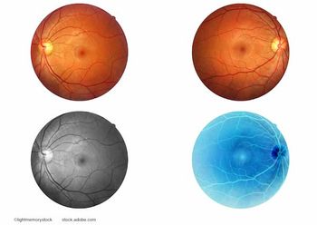
Modern imaging plays a crucial role in both diagnosis and monitoring of GA progression. The initial diagnostic workup typically includes fundus photography, fundus autofluorescence, and OCT. Although clinical trials have heavily relied on fundus autofluorescence as the gold standard for measuring disease progression and meeting FDA end points, this imaging modality presents practical challenges in routine clinical practice. The bright light required for autofluorescence imaging can be difficult for patients to tolerate, often resulting in blurry images that compromise diagnostic quality. Consequently, many clinicians obtain autofluorescence images only at baseline and every 6 to 12 months.
OCT emerges as the most practical imaging tool for routine monitoring, providing detailed cross-sectional views of atrophic areas and enabling clinicians to track progression using cursors and markers. This technology allows physicians to validate patients’ subjective experiences of visual decline by demonstrating measurable changes in atrophic lesions. OCT also helps identify important prognostic features such as hyperreflective foci, drusen volume, and markers predicting progression to complete retinal pigment epithelium atrophy. The ability to visually demonstrate disease progression to patients using OCT images serves as an important communication tool in clinical practice.
Newsletter
Keep your retina practice on the forefront—subscribe for expert analysis and emerging trends in retinal disease management.


