
|Articles|August 1, 2018
Microaneurysm turnover predicts diabetic retinopathy progression
Author(s)Laird Harrison
Micoaneurysm turnover in the early stages of diabetic retinopathy predicts how much the disease will progress, according to researchers.
Advertisement
Micoaneurysm turnover in the early stages of diabetic retinopathy predicts how much the disease will progress, according to researchers.
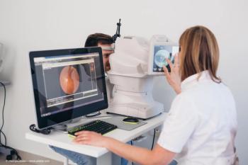
Measuring turnover is more predictive than simply counting aneurysms, and it can be done with automated analysis of just two fundus images, reported Dr Rajeev K. R. Pappuru from the L.V. Prasad Eye Institute in Hyderabad, India, and colleagues. They published the findings in the
Identifying which cases of diabetic retinopathy are most likely progress can help clinicians to prevent threats to their patients’ vision, they wrote. Systematic markers of diabetes have not proved useful for this purpose, they note. And even under similar metabolic control, some patients progress rapidly to macular oedema while others do not.
Enrollment
The researchers wanted to see whether the development of microaneurysms could provide a more useful biomarker for diabetic retinopathy progression, so they recruited 205 people with type 2 diabetes who were over 35 years of age in Coimbra, Portugal and Hyderabad, India. The patients had mild non-proliferative diabetic retinopathy defined as levels 20 to 35 according to the Early Treatment of Diabetic Retinopathy Study (ETDRS).
They had best-corrected visual acuity (BCVA) greater than 20/25 on the ETDRS chart and glycated haemoglobin (HbA1c) of 11% at most. Other criteria included no previous treatment with laser, anti-vascular endothelial growth factors or steroid intravitreal injections, and the absence of other retinal vascular disease, glaucoma, or inadequate ocular media and/or pupil dilation.
The researchers used an automated computer-aided diagnostic system, the RetmarkerDR (Retmarker), to detect microaneurysms automatically on field-two colour fundus images. Its software includes an algorithm that compares the same location in a retina between different visits. This allowed the researchers to calculate the number of microaneurysms that appeared or disappeared for each patient per time interval.
They sorted patients into three phenotypes of diabetic retinopathy progression:
• Phenotype A: low microaneurysm turnover and normal retinal thickness (MA turnover <6 and central subfield retinal thickness less than 260 µm for women or less than 275 µm for men)
• Phenotype B: low microaneurysm turnover and increased retinal thickness (MA turnover <6 and central subfield retinal thickness at least 260 µm for women or at least 275 µm for men)
• Phenotype C: high MA turnover (MA turnover ≥6) with or without increased retinal thickness.
Forty-seven patients dropped out of the study. Compared to those who stayed in the study, more of those who dropped out were Asian, and their low-density-lipoprotein cholesterol and diastolic blood pressure were higher on average.
Only 158 eyes reached central-involved macular oedema after 24 months, which was the last study visit. Twenty-seven (17.1%) progressed to more advanced ETDRS: six progressed to mild non-proliferative diabetic retinopathy (level 35), 15 to moderate non-prolific diabetic retinopathy and five to moderately severe non-proliferative diabetic retinopathy. One progressed to the high-risk proliferative form.
Of the patients in phenotype A, only 1.1% developed central-involved macular oedema, compared with 28.8% of those in phenotype B and 29.4% of those in phenotype C.
Likewise, 19.8% of phenotype A patients experienced worsening of diabetic retinopathy of at least one step. This compared with 21.7% in phenotype B and 93.3% in phenotype C.
Genetic differences
The researchers conducted a multivariate analysis to adjust for the affects of phenotypes, metabolic control and cardiovascular risk variables. They found that eyes from phenotype C had a higher risk of developing central-involved macular oedema than eyes from phenotype A (odds ratio [OD] 44.8, 95% confidence interval [CI] 6.8 to 293.8; p<0.001).
Eyes from phenotype B showed a higher chance of developing central-involved macular oedema than eyes/patients from phenotype A (OR 31.4, 95% CI 5.4 to 183.3; p<0.001). A higher percentage of the patients in India were phenotype C. And most of the eyes that progressed were from India.
Only three of the 92 eyes (3.3%) from Portugal progressed to mild non-proliferative diabetic retinopathy. Of the 57 eyes from India, 24 (42.1%) progressed to more advanced ETDRS: three to mild, 15 to moderate and five to moderately severe (level 43) non-proliferative diabetic retinopathy. One progressed to high-risk proliferative diabetic retinopathy.
The researchers noted that previous studies have showed a genetic susceptibility to diabetic retinopathy, which could explain this difference in populations. However, in general, they wrote that the study results confirmed that microaneurysm turnover rates can help identify those patients most at risk of worsening diabetic retinpathy.
Newsletter
Keep your retina practice on the forefront—subscribe for expert analysis and emerging trends in retinal disease management.
Advertisement
Related Content
Advertisement


Latest CME


Advertisement
Advertisement