Adapting medical retina service in COVID-19 era requires careful approach
Minimizing clinic visits, maximizing use of imaging modalities are key
If we have learned anything from 2020, it is this: You do not want to contract coronavirus disease 2019 (COVID-19). It is not clear if post-infection immunity to the virus lasts longer than 4 to 6 months.1 It is also increasingly apparent that many infected patients who experienced only mild symptoms actually have experienced considerable organ damage—not just to their lungs,2 but to their kidneys,3 heart,4 vasculature,5 and brain.6
Simple public health measures that are effective in reducing the virus’ transmission, such as wearing face masks in public areas, are facing resistance. It may be that the course of the COVID-19 crisis mirrors the 1918 influenza pandemic7: an initial devastating wave of cases soon followed by a far larger second wave and then a disturbingly long tail. The best form of disaster management is to hope for the best but prepare for the worst. So how does this translate into running a medical retina service?
Most patients using such a service have either age-related or diabetic eye disease, both of which are risk factors for severe disease and death from COVID-19.8 Some of these patients may be residents in residential care homes, which were hotbeds of disease activity during the last spike in cases.
This means there is a very thin tightrope to be walked: the ophthalmologist needs to see the patients as frequently as possible to maintain the patients’ vision as best as possible under these circumstances, but as infrequently as possible to minimize the risk of COVID-19 infection. How to achieve this?
Triage, sanitizer, and PPE
It starts well before the clinic. At our practice, patients are sent questionnaires 48 hours before they arrive. They are asked whether they have been in contact with a person with COVID-19, have traveled to a region with a high prevalence of COVID- 19, and if they are experiencing any COVID-19-related symptoms.
If patients are likely to have COVID-19 based on their answers, then we must decide whether seeing them in the next few days is essential or if they can wait for a 14-day “office isolation” period. We also need to decide whether to refer them to a facility that specifically deals with people who may have COVID-19 or whether we can issue them an FFP2 safety mask at their arrival, ask the office personnel to wear appropriate personal protective equipment (gown, FFP2 mask, protective eyewear), and see these patients at the end of the day.
Hand sanitizer, face masks, and social distancing are key weapons in stopping the spread of the virus: All patients are asked to sanitize and wear a mask, and the layout of the waiting area has changed, with every second seat removed to stop patients from sitting together and ensure a safe distance is maintained.
We also keep as many doors and windows open as possible to maximize the flow of fresh air in the building with the aim of reducing the concentration of any virus fomites that may be present (and to minimize the repeated touching of door handles). In larger clinics or hospitals, an outside tent can be used to perform screening procedures while ensuring adequate ventilation.
To minimize the potential for COVID-19 exposure to other patents and clinic staff, patients’ friends and family are forbidden to accompany them into the clinic. As the risk of contagion waxes and wanes, this requirement can be adjusted based on the local risk, because it is seen as a burden by many patients and their families.
The number of people in the waiting room is limited to 3 groups of 2 people. We also perform a type of scheduling triage: Patients at highest risk of getting severe COVID-19–related disease—such as those with both hypertension and type 1 diabetes—are seen in the first few appointments of the day, based on the assumption that fewer potentially infected people would have been in the clinic before them, and fomites do not survive on surfaces and in the air for a prolonged period.
Pushing the treatment interval to the max
The key to keeping treatment standards as high as possible under such difficult circumstances is chart review. If patient care can be managed remotely with a telephone call, this is better than having the patient come into the clinic.
The charts of patients receiving anti-VEGF agents to treat neovascular age-related macular degeneration or diabetic macular edema (DME) should be reviewed to see if a treat-and-extend (T&E) regimen has been pushed to the maximum. If it is feasible for a patient to visit the clinic every 3 months, we would try to push the treatment interval this long or longer.
One also needs to prepare for some difficult choices: as if the COVID-19 situation were not dire enough, the fact is that some patients may have to lose vision—a decision that may be made easier if the patient is losing vision in only 1 eye, or more difficult if the patient has only 1 remaining “good” eye. When it comes to ensuring patients retain their vision, the biggest risk of failure is loss to follow-up. Ophthalmologists must keep in contact with their patients to ensure that they attend these now-critical clinic visits for treatment that really does have to be administered in a timely manner.
It would appear that large outbreaks take about 2 to 3 months to control. For patients with DME, especially patients who are pseudophakic, use of longer-acting steroid therapy such as a dexamethasone intravitreal implant (Ozurdex, Allergan) is worth considering. When it comes to phakic patients; we have been seeing that for those with uveitis, cataracts only really start to develop after the injection of the third implant.
In countries that are still in an early wave of infections or possibly a continuous surge, longer-acting agents are even more important in reducing the number of patient visits to the clinic. The decision to use steroid treatment requires careful thought because of the potential risks of increased intraocular pressure and cataract development.
It may be less appropriate to administer steroids to younger people, but in our opinion, the ones who are really at high risk are the older individuals who have diabetes or hypertension or are obese. In the current context, it is probably better to put them on dexamethasone implant, perhaps with prophylactic antiglaucoma drops if there is a known or suspected ocular hypertension response to steroids. These patients present to the clinic less frequently and if a cataract develops, it can then be dealt with once the much-anticipated next wave of COVID-19 cases has subsided.
Why a slit lamp exam may be riskier than a surgical procedure
Cataract surgery, perhaps counterintuitively, should have a low risk of COVID-19 transmission. When performing this procedure, we test all patients for the virus 2 to 3 days before surgery; all individuals who test negative can receive the surgery.
There is a theoretical risk that the patient may have been infected in the meantime, but in the first few days, they are unlikely to transmit the virus. The patient wears a mask before, during, and after the procedure; almost all cataract procedures are performed under local anesthesia (so no intubation is required). Just before starting the surgery, patients can be asked to drop the mask so they can be given oxygen, and then they are covered with a sterile drape with a seal around the eyes.
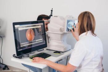
In fact, the risk during surgery versus slit lamp examinations may be lower than we believe. There is certainly less risk associated with performing OCT imaging than there is with a slit lamp exam 30 cm from the patient (even if there is a layer of Perspex screening around the slit lamp); with OCT, the ophthalmologist and the technician are sitting farther away and not directly in front of the patient.
Another noteworthy point is that wearing masks can mean lenses steam up on diagnostic instruments, and this is more likely to occur in warm and humid offices. The difference between a cold and dry day versus a hot and humid day made a 3-line difference in our patients’ performance when conducting refraction with trial lenses.
What good looks like in the new normal
If the COVID-19 pandemic worsens dramatically and is associated with a long tail, then “what good looks like” will involve assessment and treatment modalities that identify patients in need of treatment. It will extend the period between patient visits to the clinic during these critical 2- to 3-month periods. As things stand, these treatments are limited to longer-acting steroid implants and maximal T&E protocols with anti-VEGF drugs.
It is clear that in the near future some aspects of the clinic visit may be replaced by telemedicine, such as certain aspects of the regular consultation being performed over the telephone, Snellen visual acuity assessments being performed via smartphone apps, and ocular surface examinations being conducted with video consultations. However, these are clearly insufficient for monitoring the status of the retina.
Ophthalmologists need home OCT testing that patients can use to generate images, which they can then share with the retina specialist for assessment and treatment decision-making. This would dramatically reduce the number of patient visits required and ensure that the only patients who visit the clinic for treatment are truly those who need it.
References
1. Seow J, Graham C, Merrick B, et al. Longitudinal evaluation and decline of antibody responses in SARS-CoV-2 infection. medRxiv 2020.07.09.20148429. doi:10.1101/2020.07.09.20148429
2. Li H, Liu L, Zhang D, et al. SARS-CoV-2 and viral sepsis: observations and hypotheses. Lancet. 2020;395(10235):1517-1520. doi:10.1016/j. kint.2020.04.003
3. Su H, Yang M, Wan C, et al. Renal histopathological analysis of 26 postmortem findings of patients with COVID-19 in China. Kidney Int. 2020;98(1):219-227. doi:10.1016/j.kint.2020.04.003
4. Shi S, Qin M, Shen B, et al. Association of cardiac injury with mortality in hospitalized patients with COVID-19 in Wuhan, China. JAMA Cardiol. 2020;5(7):802-810. doi:10.1001/jamacardio.2020.0950
5. Cui S, Chen S, Li X, Liu S, Wang F. Prevalence of venous thromboembolism in patients with severe novel coronavirus pneumonia. J Thromb Haemost. 2020;18(6):1421-1424. doi:10.1111/jth.14830
6. Mao L, Jin H, Wang M, et al. Neurologic manifestations of hospitalized patients with coronavirus disease 2019 in Wuhan, China. JAMA Neurol. 2020;77(6):683-690. doi:10.1001/jamaneurol.2020.1127
7. 1918 pandemic Influenza: three waves. Centers for Disease Control and Prevention. May 11, 2018. Accessed July 27, 2020. https://www.cdc.gov/flu/pandemic-resources/1918-commemoration/three-waves.htm
8. Jordan RE, Adab P, Cheng KK. Covid-19: risk factors for severe disease and death. BMJ. 2020;368:m1198. doi:10.1136/bmj.m1198
Marc D. de Smet, MD, PhD
e: mddesmet1@mac.com
De Smet is director of MIOS in Lausanne, Switzerland (www.retina-uveitis.eu ), and medical director of Preceyes Medical Robotics (www.preceyes.nl ) in Eindhoven, The Netherlands. He has no financial disclosures related to the content of this article.
Marco Mura, MD
p: 410-955-2777
Mura is chief of the Retina Division at King Khaled Eye Specialist Hospital in Riyadh, Saudi Arabia, and an associate professor of ophthalmology at the Wilmer Eye Institute, Johns Hopkins University School of Medicine in Baltimore, Maryland. Mura has no financial interests in the subject matter.
Newsletter
Keep your retina practice on the forefront—subscribe for expert analysis and emerging trends in retinal disease management.


