


Investigators find imaging method may be useful to monitor disease progression.
Investigators find imaging method may be useful to monitor disease progression.
Novel treatments may be on the horizon for the advanced form of age-related macular degeneration.
Option detects treatment-naive nonexudative macular neovascularization in eyes with dry age-related macular degeneration.

Mark Breazzano, MD, discusses the future of telehealth for retina specialists.
A team of investigators found that the retina may offer signs of COVID-19 infection before symptoms present.
Dual imaging may reduce costs, unnecessary referrals, telehealth study results show.
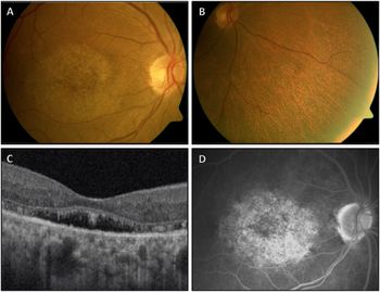
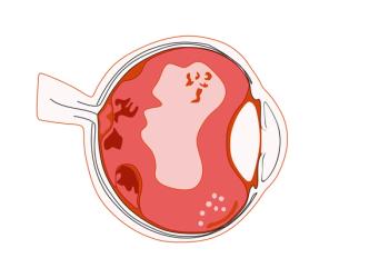
Structural photoreceptor abnormalities and an area of central sparing found.
Study of subjects with rare inherited form of AD shows increased retinal capillary perfusion during the presymptomatic stage.
Deep learning predictions of the retinal nerve fiber layer thickness based solely on fundus photographs can be used to monitor the risk of future glaucomatous conversion in glaucoma suspects.
Investigators corroborate the hypothesis that retinal ganglion cells with dendrites stratified in the off sublaminae could be damaged.

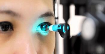
According to investigators, the new technology is designed to detect telltale signs of major blinding diseases in retinal blood and tissue that typically go unseen until it is too late.
Measures of inflammation can predict treatment outcomes for patients

Retinal imaging tests are providing material to train and test decision support systems.

Michael Mbagwu, MD, speaks on the highlights of his virtual 2021 ARVO presentation regarding the development of an algorithm tool that links patient imaging to their clinical data, specifically within the AAO IRIS Registry.
Study evaluates the impact of surgical procedure on visual outcomes in patients

Easier, more accessible functional studies have been made possible

Technologies offer different approaches to different scenarios

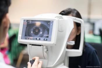
Worldwide increase in technologies for ophthalmic use is dramatic

For ophthalmologists, the interactive landscape of surgical visualization, including 3D digital surgery, robotics, and an OCT-enriched surgical theater, is leading to new opportunities.

Short FLIO flecks may appear before changes are seen on other imaging modalities, including OCT.
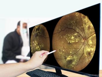
High-quality images can be useful for early diagnosis, prognosis, monitoring

Results of a prospective trial show the tool is ready for clinical use and potential deployment for remote telemedical use.

Study explores correlations between functional and structural tests
Investigators tested the hypothesis that changes in certain areas of the macular are more relevant to AMD. What did their findings reveal?
The Audacious Goals Initiative of the National Eye Institute is focused on developing noninvasive imaging advances to help patients with vision loss.